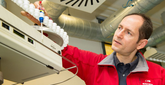
Heiko Möller works at the interface of chemistry, biology, and medicine, which makes his research special. “We are interested in biomolecules,” says the Professor for Analytical Chemistry. These include complex protein molecules, like enzymes and antibodies. Nearly every function of biomolecules depends on their reaction with binding partners. Findings on the chemical principles of these interactions can be used to develop medicine or vaccines.
Potsdam chemists generally examine proteins produced in bacteria. As structural biologists, Möller and his team are interested in high-resolution 3D protein structures – particularly in which partners they interact with, what changes such interactions entail, and how they are regulated. Their approach involves combining NMR spectroscopy with other experimental and theoretical methods.
Basic research technique – NMR spectroscopy
X-ray crystallography is the tool most frequently used to solve 3D protein structures. About 90% of high-resolution 3D structures are determined by this method. Some proteins, however, do not crystallize and the method is not usable. Möller is interested in one such protein group, whose structures are normally made visible through nuclear magnetic resonance spectroscopy. “NMR spectroscopy is a basic research technique for chemists, which has become essential in laboratory practice,” Möller explains. This method allows molecular structures to be analyzed down to the smallest detail. The researchers determine which atoms in a molecule are linked as well as the 3D arrangement of functional groups, for example in a sugar molecule. NMR spectroscopy allows for the verification of substance purity. For the current project “Structural Biology of Focal Adhesion: Cell Adhesion and Migration”, researchers of the University of Potsdam are collaborating with colleagues from the University of Konstanz, where Möller worked before coming to Potsdam.
Protein Paxillin
“When we realized that ‘our’ protein would not crystallize under any condition tested, we turned to NMR spectroscopy,” the chemist explains the approach. This, however, requires relatively large quantities of stable protein. Whereas many biological experiments work with only a few micrograms, structural analysis by NMR requires about 10 milligrams (i.e. 10,000 micrograms). The fact that the proteins must neither change nor decompose presented a problem. The so-called optimization of the protein construct, i.e. the format necessary to continue the research, may well take a few years. A “well behaved” protein preparation is a prerequisite for structural examination.
Proteins have to be labeled with isotopes before NMR spectroscopy can analyze their structures. Since they are usually only available in small amounts, 15N-ammonium chloride, 13C-glucose, and some vitamins and minerals have to be “fed” to the protein-producing bacteria. Under these conditions, isotopes accumulate in the bacteria and produce a large number of appropriately labeled proteins. “We then clean the bacteria off of the proteins by harvesting the cells, destroying the cell membrane, and isolating the desired protein – usually in several steps – from the cytoplasm, the liquid inside of the cell,” Möller says.
The researchers are determining the 3D structures of segments of the relatively large paxillin protein. This includes determining the NMR-spectroscopic resonance frequencies of hydrogen, nitrogen, and carbon nuclei. It is a puzzle that can take many weeks or even months to solve, followed by complex calculation methods to determine the structure.
Möller is aware that his research “only” looks at a tiny part of reality. “That, however, spurs my scientific mind,” he says. For the current project this means practical application, like medication, is quite unlikely for the coming years. Research needs time and perseverance.
Paxillin plays a significant role in cell migration. Cells do not move like humans with bones and muscles but by protrusion. Filaments adhere to the surface and the trailing cell edge is retracted. The process – comparable to the locomotion of snails and worms – is continuously repeated. Cells adhere to surfaces, other cells, or tissues. They brachiate or move through something. Immune cells – like leukocytes – that move into inflamed tissue are a typical example. This also applies to cancer cells that move from the tumor to other parts of the body and form metastases. Thus, cell migration – also significant for vital processes like growth, development, and differentiation – also influences pathological processes like cancer and nerve cell degeneration.
The Researcher
Prof. Heiko M. Möller studied chemistry in Hamburg. Since 2014 he has been Professor for Analytical Chemistry at the Institute of Chemistry at the University of Potsdam.
Contact
Universität Potsdam
Institut für Chemie
Karl-Liebknecht-Straße 24-25, 14476 Potsdam
E-Mail: heiko.moelleruuni-potsdampde
The Project
Structural Biology of Focal Adhesion: Cell Adhesion and MigrationParticipating: Prof. Heiko M. Möller, University of Potsdam – Collaboration with Prof. Christof Hauck, University of Konstanz; funded as part of the Collaborative Research Center (SFB) 969 “Proteostasis”.Duration: 2016–2019 (2nd funding period)
Text: Dr. Barbara Eckardt
Translation: Susanne Voigt
Published online by: Agnetha Lang
Contact for the editorial office: onlineredaktionuuni-potsdampde

