Zum Hauptinhalt springen

Verwendete Methoden

Spektroskopie

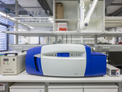
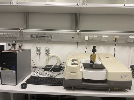
CD-Spektrometer im Labor
Foto: Thomas Roese
Applied Photophysics Chirascan

CD-Spektroskopie

Strukturaufklärung  von chiralen (Bio)Molekülen und Untersuchung ihrer Wechselwirkungen mit anderen Molekülen


CD-Spektrometer im Labor
Foto: Thomas Roese
Applied Photophysics Chirascan
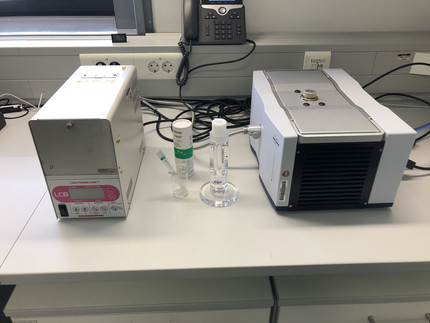
Gerät für Elektronspinresonanzmessungen
Foto: Beatrice Battistella
Bruker Magnettech ESR5000

EPR-Spektroskopie

Elektronenspinresonanz-Messungen paramagnetischer Substanzen


Gerät für Elektronspinresonanzmessungen
Foto: Beatrice Battistella
Bruker Magnettech ESR5000
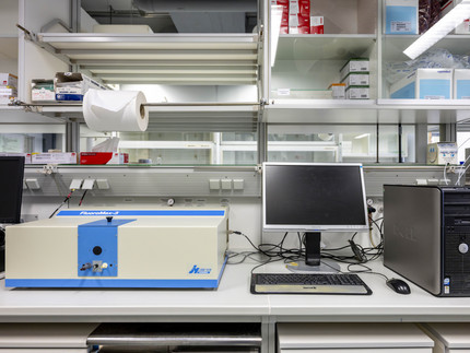
Fluorimeter FluoroMax im Labor
Foto: Thomas Roese
Horiba Scientific FluoroMax 2

Fluoreszenzspektroskopie

Fluorimetrische Messungen


Fluorimeter FluoroMax im Labor
Foto: Thomas Roese
Horiba Scientific FluoroMax 2
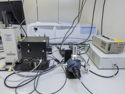
Anlage zur Bestimmung der Fluoreszenquantenausbeute
Foto: Thomas Roese
Hamamatsu PL C9920-2

Fluoreszenz-Quantenausbeute

Bestimmung der Quantenausbeute im Bereich 300-950 nm


Anlage zur Bestimmung der Fluoreszenquantenausbeute
Foto: Thomas Roese
Hamamatsu PL C9920-2
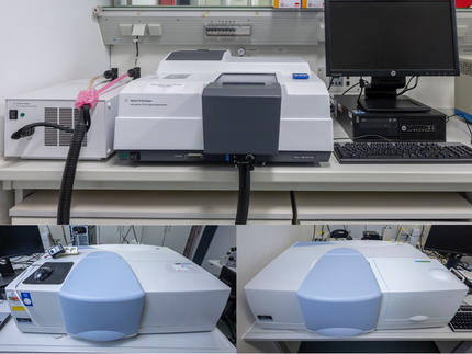
Drei UV/VIS-Spektrometer
Foto: Thomas Roese
Agilent Varian Cary 100 Bio UV/VIS spectrophotometer (oben), Perkin Elmer Lambda 750 und 950 (unten links bzw. rechts)

UV/VIS-Spektroskopie

Messung der Absorption im Bereich 190–900 nm (Flüssigkeiten und Feststoffe), temperaturabhängige Messungen mit Sechsfachküvettenwechsler


Drei UV/VIS-Spektrometer
Foto: Thomas Roese
Agilent Varian Cary 100 Bio UV/VIS spectrophotometer (oben), Perkin Elmer Lambda 750 und 950 (unten links bzw. rechts)

Elektrochemie

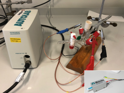
Metrohm DropSens μStat 400 Bipotentiostat/Galvanostat
Foto: Thomas Roese
Metrohm DropSens μStat 400 Bipotentiostat/Galvanostat

Cyclovoltammetrie

Bestimmung von Redoxpotentialen (mit Chipelektroden)


Metrohm DropSens μStat 400 Bipotentiostat/Galvanostat
Foto: Thomas Roese
Metrohm DropSens μStat 400 Bipotentiostat/Galvanostat
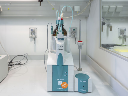
Potentiometrie-Aufbau im Labor
Foto: Thomas Roese
Metrohm 888 Titrando

Potentiometrie

Potentiometrische pH-Titrationen


Potentiometrie-Aufbau im Labor
Foto: Thomas Roese
Metrohm 888 Titrando
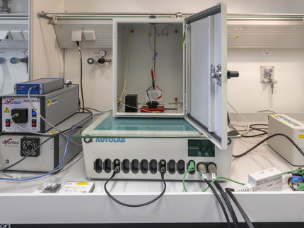
Anlage für spektroelektrochemische Messungen
Foto: Thomas Roese
Photometrie-Setup (UV/VIS-Deuterium-Halogenquelle und NIR gekoppelt mit Avantes SensLine-Detektor) und Metrohm-Potentiostat Autolab PGSTAT101 und/oder Autolab PGSTAT302N

Spektroelektrochemie

Simultane Bestimmung des Redoxpotentials und der Absorption im UV/VIS/NIR-Bereich

Anlage für spektroelektrochemische Messungen
Foto: Thomas Roese
Photometrie-Setup (UV/VIS-Deuterium-Halogenquelle und NIR gekoppelt mit Avantes SensLine-Detektor) und Metrohm-Potentiostat Autolab PGSTAT101 und/oder Autolab PGSTAT302N

Elementanalytik

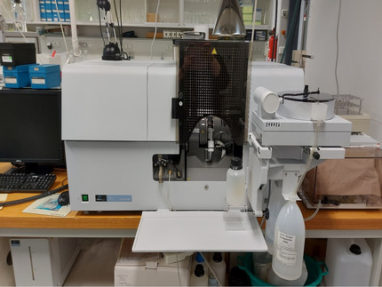
AAS-Gerät im Labor
Foto: Susanne Lubahn
Perkin Elmer AAnalyst800

AAS

Atomabsorption mittels Flammen-, Graphitrohr- und Hydridtechnik zur Analyse von Metallen und Halbmetallen


AAS-Gerät im Labor
Foto: Susanne Lubahn
Perkin Elmer AAnalyst800
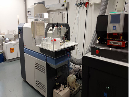
ICP-MS-Gerät im Labor
Foto: Susanne Lubahn
Thermo Fisher Element 2

ICP-MS

Gehaltsbestimmung von Elementen mittels doppelt fokussierendem hochauflösendem Massenspektrometer (nicht Halogene, Edelgase, Kohlenstoff, Stickstoff, Wasserstoff, Sauerstoff)


ICP-MS-Gerät im Labor
Foto: Susanne Lubahn
Thermo Fisher Element 2
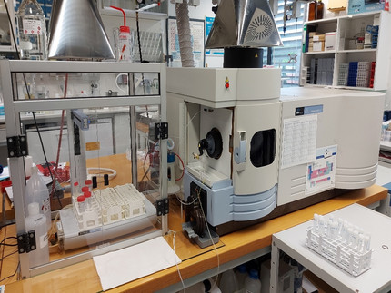
ICP-OES-Gerät im Labor
Foto: Susanne Lubahn
Perkin Elmer Optima 5300DV

ICP-OES

Gehaltsbestimmung von Elementen mittels Atom- und Ionenemission (außer Halogene, Edelgase, Kohlenstoff, Stickstoff, Wasserstoff, Sauerstoff)


ICP-OES-Gerät im Labor
Foto: Susanne Lubahn
Perkin Elmer Optima 5300DV
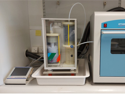
Aufschlussmikrowelle im Labor
Foto: Susanne Lubahn
MLS Ethos 1

Aufschlussmikrowelle

saure und basische Aufschlüsse von festen Stoffen


Aufschlussmikrowelle im Labor
Foto: Susanne Lubahn
MLS Ethos 1
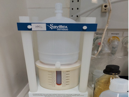
Säuredestillationsanlage im Labor
Foto: Susanne Lubahn
Savillex DST1000

Säuredestillation

Säureaufreinigung nach dem Subboiling-Prinzip


Säuredestillationsanlage im Labor
Foto: Susanne Lubahn
Savillex DST1000

Sonstiges

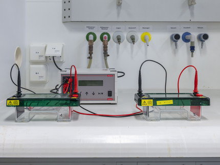
Elektrophorese-Kammern für Agarose-Gele in einem Abzug
Foto: Thomas Roese
Roth Rotiphorese PROfessional I, nicht auf dem Bild: Bio-Rad Mini-PROTEAN Tetra Cell

Gelelektrophorese

Auftrennung und Analyse von Protein(fragment)en (vertikal, PAGE) und DNA (horizontal, Agarose)


Elektrophorese-Kammern für Agarose-Gele in einem Abzug
Foto: Thomas Roese
Roth Rotiphorese PROfessional I, nicht auf dem Bild: Bio-Rad Mini-PROTEAN Tetra Cell
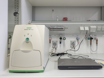
Gelscanner
Foto: Thomas Roese
Bio-Rad Gel Doc EZ Imager

Gelscanner

Visualisierung und Quantifizierung von Gelelektrophorese-Experimenten mit verschiedenen Einsätzen (Trays) je nach Gelfärbe-Methode:

  • UV Sample Tray (für Bestrahlung mit UV-Licht)
  • Stain-free Sample Tray (für ungefärbte Gele)
  • White Sample Tray (Coomassie Blau-, Kupfer-, Silber-, Zink-Färbung)
Gelscanner
Foto: Thomas Roese
Bio-Rad Gel Doc EZ Imager
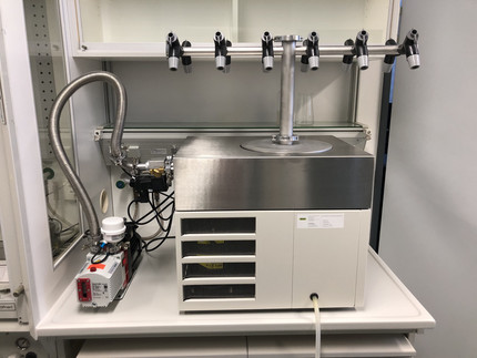
Lyophilisator
Foto: Julian Heinrich
Büchi Lyovapor L-200

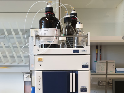
HPLC

Aufreinigung (semi-präparativ, max. 50 mg) von organischen Verbindungen insbesondere Peptiden, Reinheitsbestimmung (analytisch)


Lyophilisator
Foto: Julian Heinrich
Büchi Lyovapor L-200
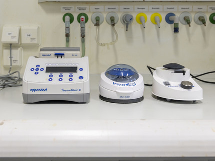
Inkubator, Minizentrifuge, Vortexer
Foto: Thomas Roese
Eppendorf ThermoMixer C, Minizentrifuge, Vortexer

Inkubieren und Mischen

Inkubation von Lösungen in Eppendorf-Gefäßen (0,5 und 1,5 mL) im Temperaturbereich 1–100 °C bei max. 3000 rpm, Zentrifugieren und Vortexen


Inkubator, Minizentrifuge, Vortexer
Foto: Thomas Roese
Eppendorf ThermoMixer C, Minizentrifuge, Vortexer
Lyophilisator mit Pumpe
Foto: Julian Heinrich
Büchi Lyovapor L-200

Lyophilisator

Gefriertrocknung von wässrigen Proben


Lyophilisator mit Pumpe
Foto: Julian Heinrich
Büchi Lyovapor L-200
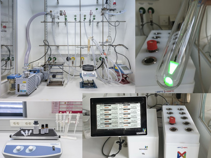
Abzug mit Vakuum-/Inertgasline, Pumpe, Magnetrührer sowie Schüttler mit Spritzen und Reagenzien für die Festphasenpeptidsynthese
Foto: Thomas Roese
Aufbau für Synthesechemie unter Inertgas (oben), Materialien und Reagenzien für die manuelle Festphasenpeptidsynthese (unten)

Synthese

  • Organische Synthese und Koordinationschemie, teilweise unter Inertgasatmosphäre
  • Festphasenpeptidsynthese und Biokonjugatchemie
Abzug mit Vakuum-/Inertgasline, Pumpe, Magnetrührer sowie Schüttler mit Spritzen und Reagenzien für die Festphasenpeptidsynthese
Foto: Thomas Roese
Aufbau für Synthesechemie unter Inertgas (oben), Materialien und Reagenzien für die manuelle Festphasenpeptidsynthese (unten)

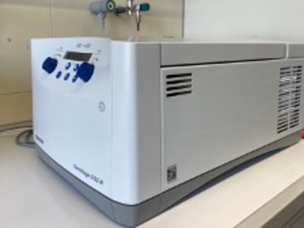
Zentrifuge

Zentrifugation von Falcon-Tubes (15 und 50 mL) und Eppendorf-Gefäßen (1,5 und 2 mL) im Temperaturbereich -9 bis 40 °C bei max. 4400 rpm